
Vol. 2 No. 3 (2025)
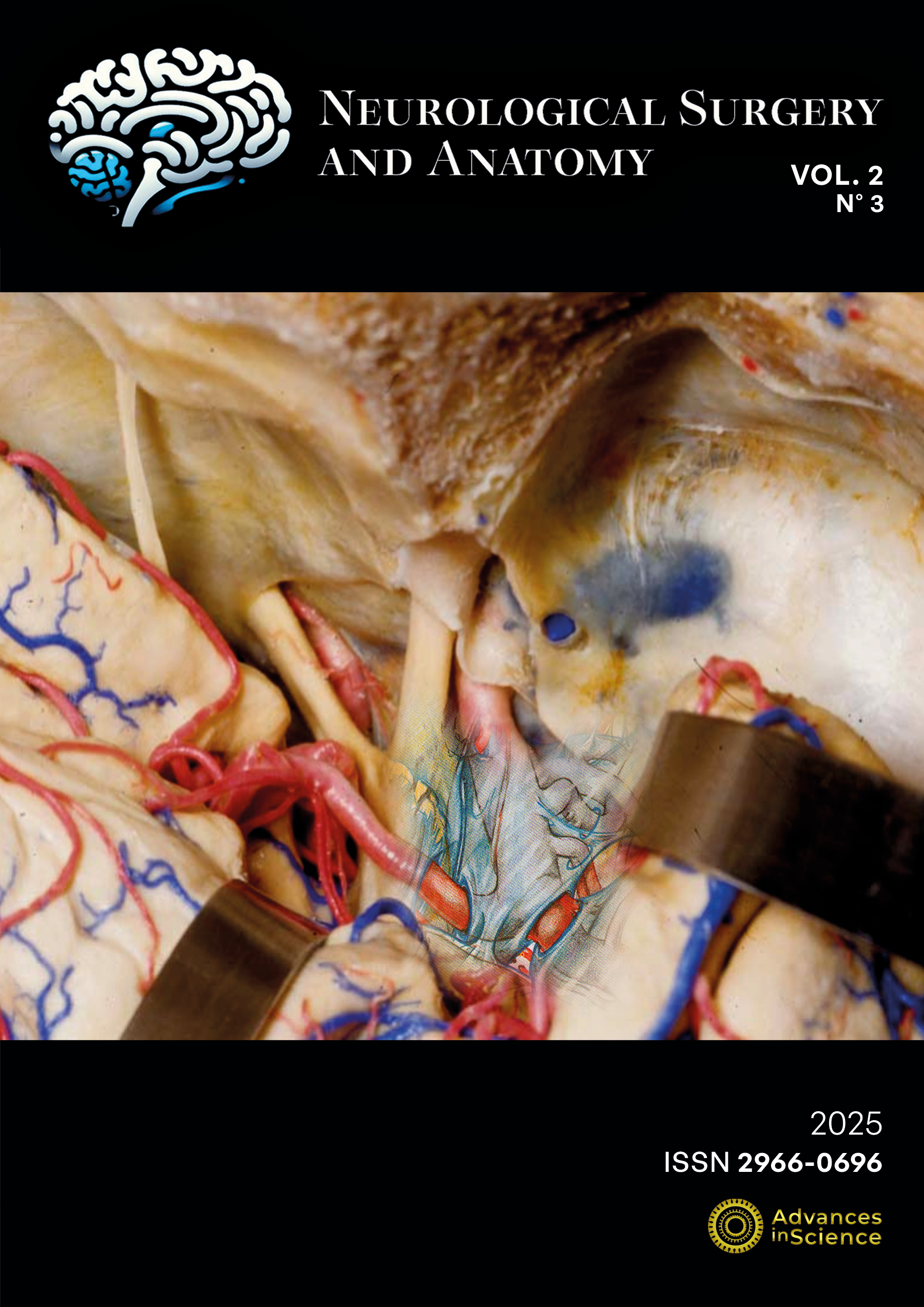
The cover of this issue features a microsurgical dissection of a cadaveric injected specimen, simulating a pterional, transylvian approach to the carotid bifurcation. Superimposed on this image is an artistic rendering of the arachnoidal layers, adapted from a drawing by Mr. Peter Roth, medical illustrator to Prof. Yasargil.
This composition illustrates the original contribution of Vasconcelos Jr. et al., exploring the anatomical limits of the sylvian cistern. The historical context behind this cover is further enriched by the contribution of Cavalcanti de A. Martins, offering readers an additional perspective on this giant of Medical Illustration.
Published:
2025-12-30