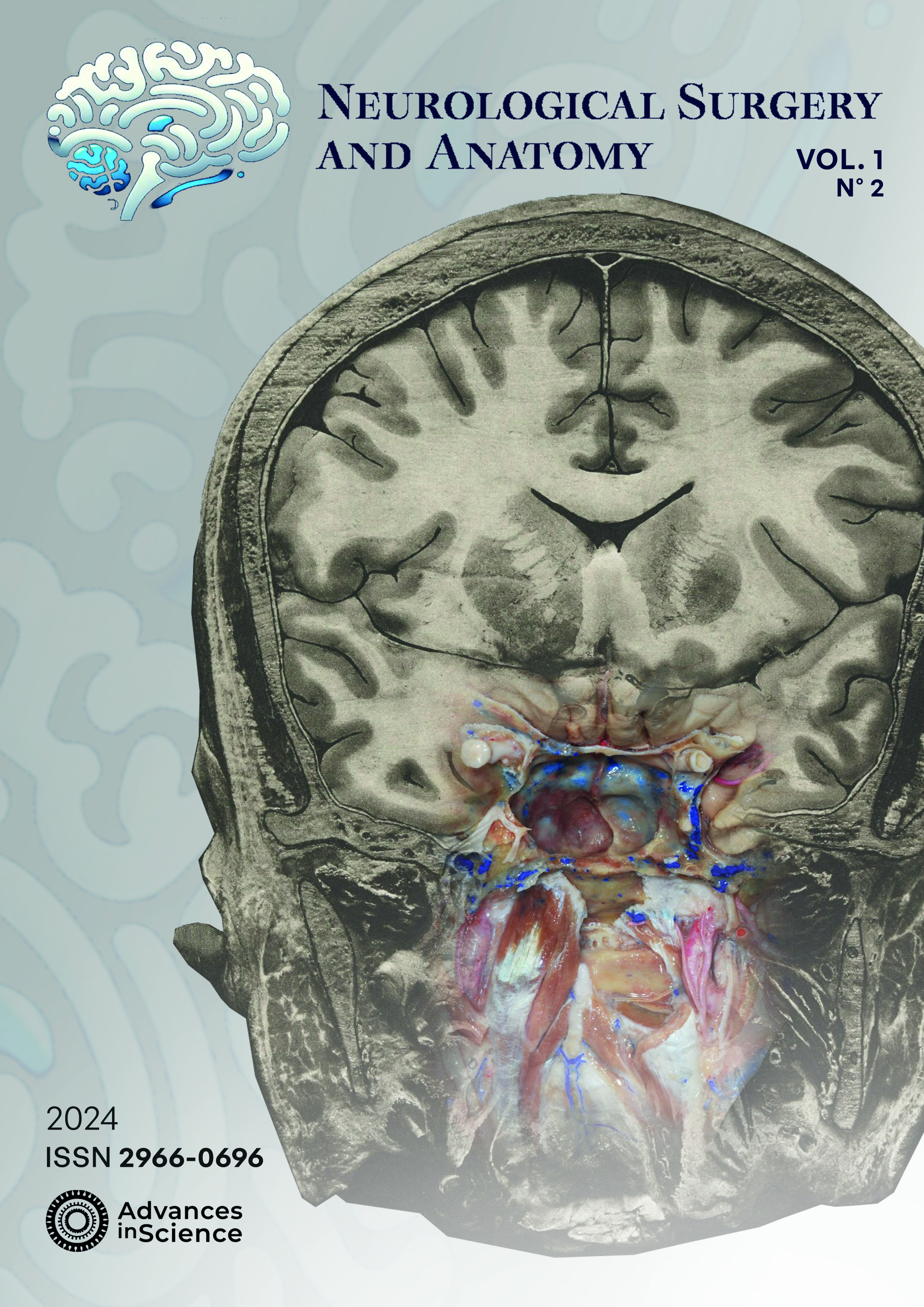
Problem based neuroanatomy of cranial nerves: case of nerve intermedius
DOI:
https://doi.org/10.37085/nsa.2024.14Keywords:
Cranial Neves, Medical Schools, Neuroanatomy, Cadaver , Human , Students, Medical StudentsAbstract
Introduction
Neuroanatomy taught at Medical School needs to be clinically meaningful and guide future acquisition of knowledge helpful in diagnosing and treating patients. Problem-based anatomy has been used to help bridge the gap between knowledge acquired in the lab and the requirements needed for daily medical practice.
Objective
This study evaluates the inception of data from Anatomage© Table combined with microsurgical dissection of cadaveric human injected specimens, as auxiliary to acquisition of anatomical milestones required for the cranial nerves, for medical students.
Methods
The study was conducted at Medical School of Pernambuco, Brazil. The Anatomage Table 10.0 was browsed for clinical cases, whose images would illustrate potential or overt pathological involvement of at least one segment of a cranial nerve. Each case was combined with microsurgical anatomy of the region to build an independent, question-lead, educational content, clarifying the anatomical milestones required to interpret, evaluate and treat similar patients.
Results
Seventeen illustrative clinical cases were selected for this purpose among 52 in-built cases at this version of the anatomical table. The inclusion of clinical cases brought a new appeal for the cranial nerve content, since it could be included both at graduation or post-graduate levels. Beyond signaling the continuous, individual process of learning anatomy, it also offers support beyond the lab walls, for the student on his/her individual learning journey.
Conclusion
This study displays the potential of technological tools, when combined with other resources, namely microsurgical dissections, to allow for creation of new and clinically significant learning resources.
References
Alasmari WA. Medical Students’ Feedback of Applying the Virtual Dissection Table (Anatomage) in Learning Anatomy: A Cross-sectional Descriptive Study. Adv Med Educ Pract. 2021 Nov;Volume 12:1303–7. Doi: 10.2147/AMEP.S324520
Gray H, Collins P. Gray’s Anatomy: The Anatomical Basis of Clinical Practice. 39th ed. Standring S, Harold E, Berkovitz BKB, editors. Elsevier Churchill Livingstone; 2005. 1627 p.
Porras‐Gallo MI, Peña‐Meliáan Á, Viejo F, Hernáandez T, Puelles E, Echevarria D, et al. Overview of the History of the Cranial Nerves: From Galen to the 21st Century. Anat Rec. 2019 Mar 9;302(3):381–93. Doi: 10.1002/ar.23928
Martins C, Martins A, Monteiro C, Tenório D, Lima D. A Fábrica do Corpo: O Sulco Limitante. 2023. [Internet]. Spotify: FPS Podcast; 2023 Oct [cited 2024 May 29]. Available from: https://podcasters.spotify.com/pod/show/fps-podcast/episodes/A-Fbrica-do-Corpo-01--Sulco-Limitante-e2baj6u
Puelles L, Tvrdik P, Martínez‐de‐la‐torre M. The Postmigratory Alar Topography of Visceral Cranial Nerve Efferents Challenges the Classical Model of Hindbrain Columns. Anat Rec. 2019 Mar 2;302(3):485–504. Doi: 10.1002/ar.23830
Alfieri A, Strauss C, Prell J, Peschke E. History of the nervus intermedius of Wrisberg. Annals of Anatomy - Anatomischer Anzeiger. 2010 May;192(3):139–44. Doi: 10.1016/j.aanat.2010.02.004
Alfieri A, Fleischhammer J, Prell J. The Functions of the Nervus Intermedius. American Journal of Neuroradiology. 2011 Aug;32(7):E144–E144. Doi: 10.3174/ajnr.A2624
Martins C, Campero Á, Yasuda A, Valença M, Rhoton Jr. AL. Eminência arqueada. Relações topográficas e importância cirúrgica. JornalBrasileirodeNeurocirurgia. 2004;15(1):11–6.
Rhoton AL, Kobayashi S, Hollinshead WH. Nervus Intermedius. J Neurosurg. 1968 Dec;29(6):609–18. Doi: 10.3171/jns.1968.29.6.0609
Sachs E. The Role of the Nervus Intermedius in Facial Neuralgia. J Neurosurg. 1968 Jan;28(1):54–60. Doi: 10.3171/jns.1968.28.1.0054
Bischoff EPE. Mikroskopische Analyse der Anastomosen der Kopfnerven [Internet]. Munchen; 1865 [cited 2024 Sep 1]. Available from: https://wellcomecollection.org/works/vftsqcxj
Burmeister HP, Baltzer PA, Dietzel M, Krumbein I, Bitter T, Schrott-Fischer A, et al. Identification of the Nervus Intermedius Using 3T MR Imaging. American Journal of Neuroradiology. 2011 Mar;32(3):460–4. Doi: 10.3174/ajnr.A2338
Scheller C, Rachinger J, Prell J, Kornhuber M, Strauss C. Schwannoma of the intermediate nerve. J Neurosurg. 2008 Jul;109(1):144–8. Doi: 10.3171/JNS/2008/109/7/0144
Møller A. Vascular compression of cranial nerves. I. History of the microvascular decompression operation. Neurol Res. 1998 Dec 20;20(8):727–31. Doi: 10.1080/01616412.1998.11740591
Alfieri A, Fleischhammer J, Strauss C, Peschke E. The central myelin–peripheral myelin transitional zone of the nervus intermedius and its implications for microsurgery in the cerebellopontine angle. Clinical Anatomy. 2012 Oct 20;25(7):882–8. Doi: 10.1002/ca.22025
Tubbs RS, Steck DT, Mortazavi MM, Cohen-Gadol AA. The Nervus Intermedius: A Review of Its Anatomy, Function, Pathology, and Role in Neurosurgery. World Neurosurg. 2013 May;79(5–6):763–7. Doi: 10.1016/j.wneu.2012.03.023
Storey CE. Then there were 12: The illustrated cranial nerves from Vesalius to Soemmerring. J Hist Neurosci. 2022 Jul 3;31(2–3):262–78. Doi: 10.1080/0964704X.2022.2033077
Hildebrand R. Soemmerring’s work on the nervous system: a view on brain structure and function from the late eighteenth century. Anat Embryol (Berl). 2005 Dec 5;210(5–6):337–42. Doi: 10.1007/s00429-005-0027-3
Bork F, Stratmann L, Enssle S, Eck U, Navab N, Waschke J, et al. The Benefits of an Augmented Reality Magic Mirror System for Integrated Radiology Teaching in Gross Anatomy. Anat Sci Educ. 2019 Nov 19;12(6):585–98. Doi: 10.1002/ase.1864
Bergman EM, de Bruin AB, Herrler A, Verheijen IW, Scherpbier AJ, van der Vleuten CP. Students’ perceptions of anatomy across the undergraduate problem-based learning medical curriculum: a phenomenographical study. BMC Med Educ. 2013 Dec 19;13(1):152. Doi: 10.1186/1472-6920-13-152


